The search results described on this page were published in the following article:
- “The use of metaverse in fetal medicine and gynecology“
European Journal of Radiology
experiment 1
Remote presentation where the instructor presents a case to participants who have joined Virtual Reality from different locations. The group is immersed in a virtual room and has as study material the digital medical data generated by various processes.
Multidisciplinary discussion in the metaverse of a 28-week fetus with type II sacrococcygeal teratoma. The 3D reconstruction with segmentation of the fetal organs with colored labels was from a fetal MRI.
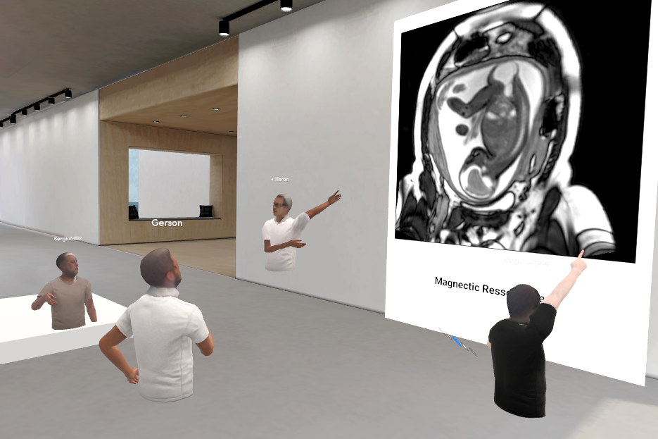
Participants view the result of an MRI scan that can be interactively manipulated to navigate 2D slices of 3D data
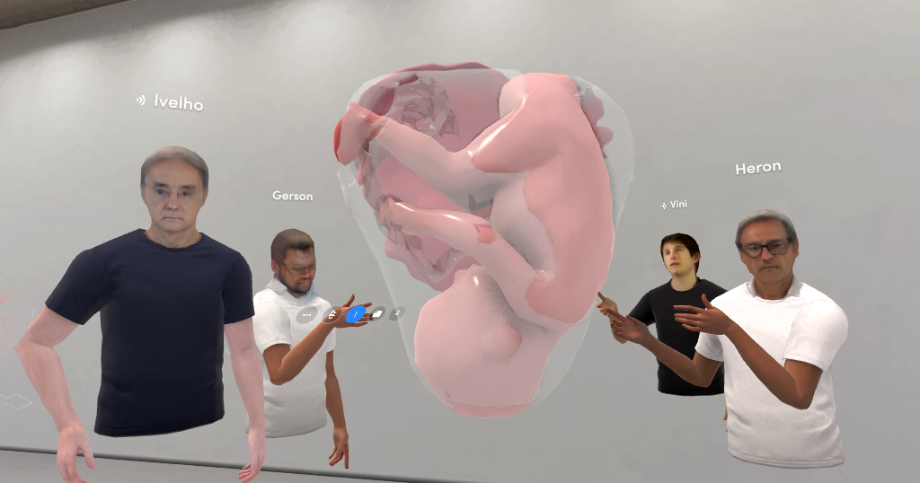
The instructor shows a 3D reconstruction of a real fetus to the class. Note that the virtual object can be spatially inspected freely with 6 degrees of freedom (translation and rotation) plus scaling.
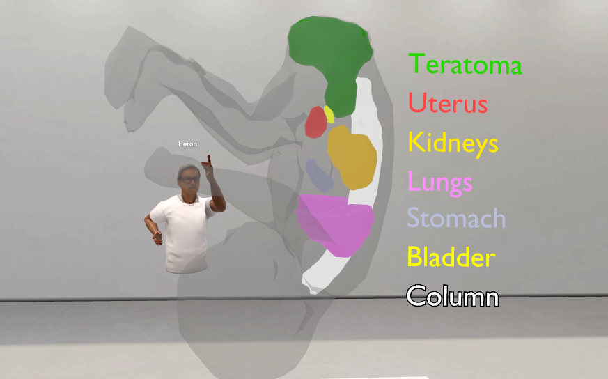
The instructor presents a 3D virtual model with segmentation of the fetal organs to discuss various anatomical aspects.
experiment 2
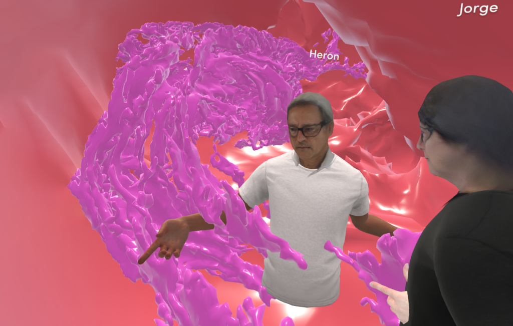
Demonstration of the possibility of having a shared meeting virtually within the 3D structure of a fallopian tube that has been digitized by micro CT in high resolution